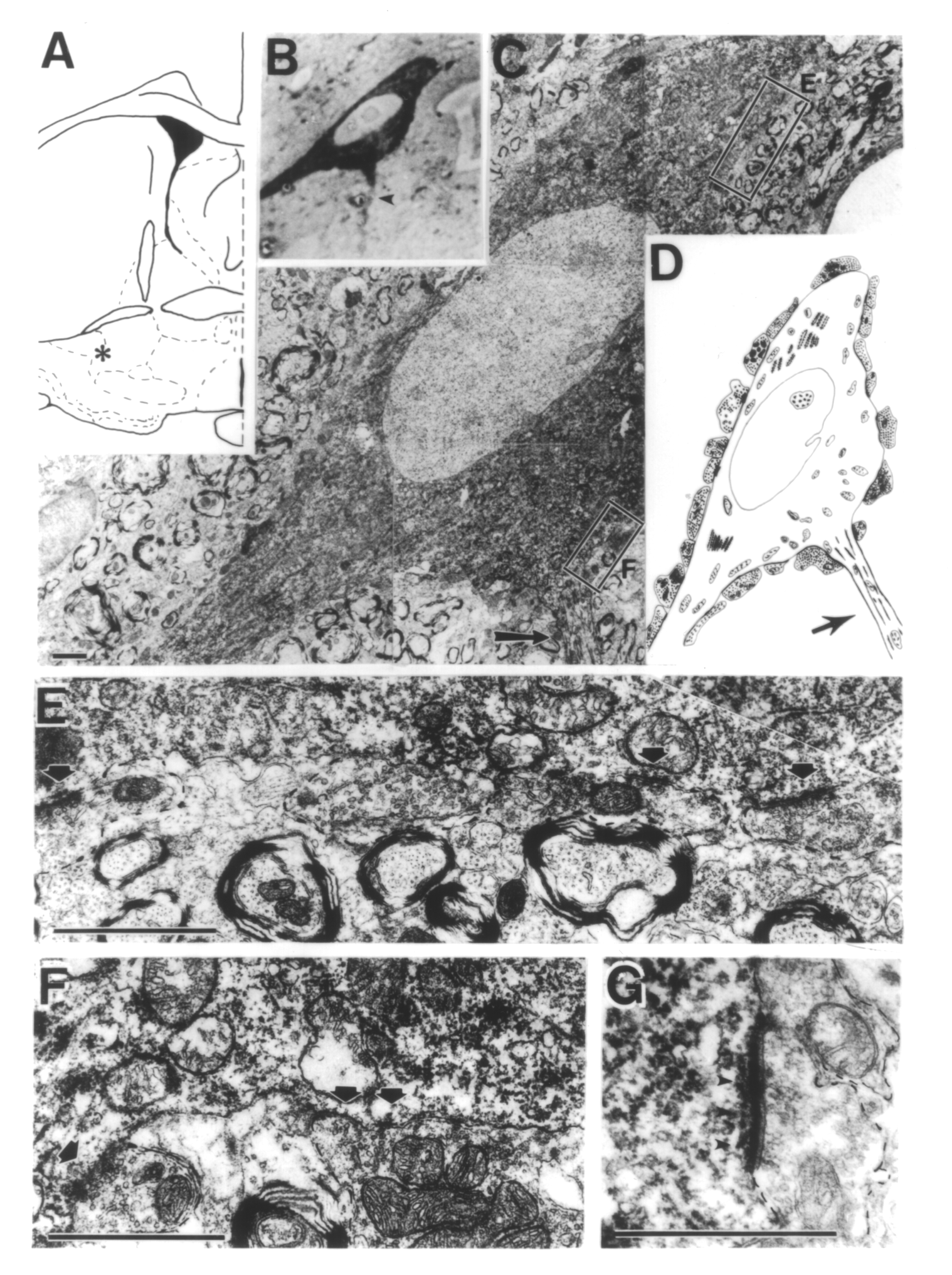
Figure 1.12.1.2. Electron micrographs showing several synapses on the cell body of a cholinergic neuron located near the substriatal gray (asterisk in A). B Appearance of the cholinergic neuron in a semithin section. Arrows in Figs. (B-D) point to the initial segment of the axon. C Low magnification electron micrograph of the perikaryon. Boxed areas are enlarged in (E) and (F). D Schematic diagram showing the distribution of boutons around the cell body. Except one terminal (arrowhead) all of them showed in this or adjacent sections symmetric synapses. E Enlarged view of the upper box in (C). Four boutons are seen in this field, three of them are in synaptic contacts (arrows) with the perikaryon. F Enlarged view of the lower box in (C) showing the axon hillock area. Both terminals enter symmetric synaptic contacts with this neuron. G Shows an asymmetric axosomatic synapse with prominent subjunctional bodies (arrowheads) from a different plane of section. Scale: C: 2 ”m; E-G 1 ”m.