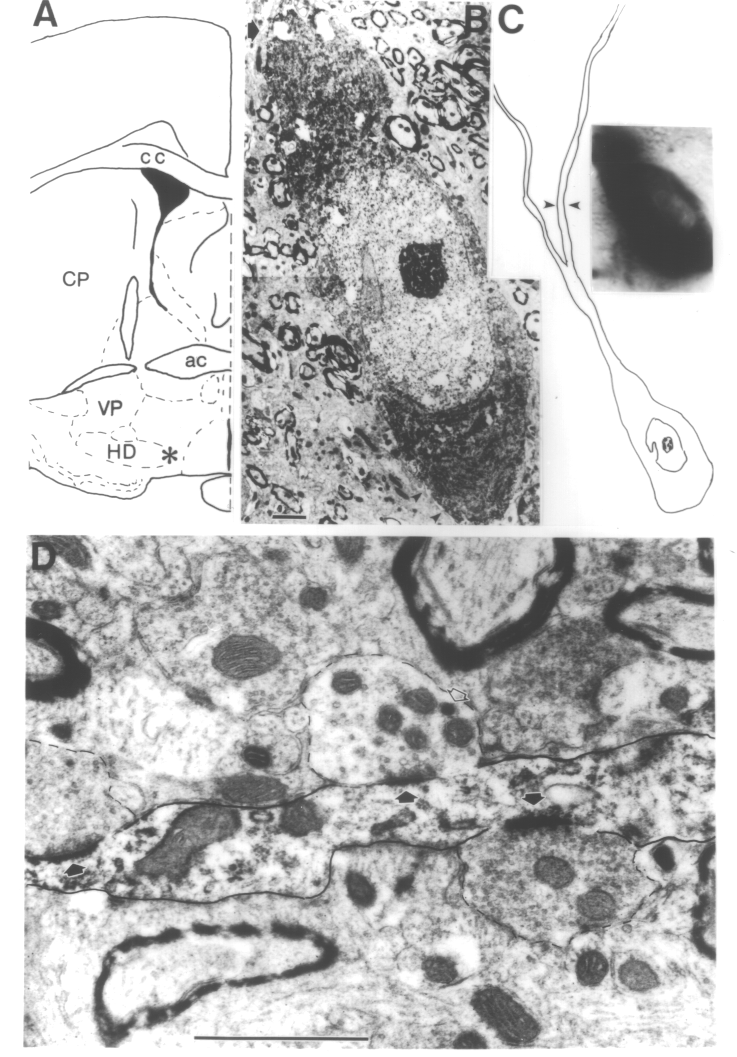
Figure 1.12.1.1. Electronmicrographs showing a cholinergic cell body (B) and a portion of its dendrite (D). A Asterisk indicates the location of the neuron, medial to the horizontal limb of the diagonal band (HD). CP, nucleus caudate putamen; VP, ventral pallidum; ac, anterior commissure; cc, corpus callosum; B Electron micrograph of the cell body. Large part of the plasmalemmal surface is covered by glial processes (arrowheads at lower right). At this plane only one synapse (arrow at upper left) is recognizable. C Schematic drawing of the neuron. Arrowheads denote the portion of the dendrite shown in (D). Insert is a photomicrograph from the cell body. D Electron micrograph showing several boutons in close associations (synapses are indicated by arrows) with this dendrite. Open arrow points to a large dense core vesicle reminiscent of a neurophysin-containing neurosecretory granule. Scale: B: 2 µm; D: 1 µm.